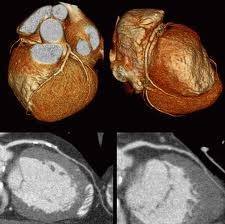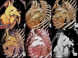
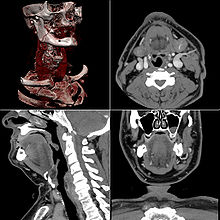
In order to maximize the diagnostic accuracy of our CT studies, we routinely acquire our images at the narrowest slice thickness appropriate to the study. Generally this will be 1 mm for chest, abdomen and pelvis, and 0.5 mm for head, neck and distal extremities. This results in a large number of axial images, numbering in the hundreds and in some cases over a thousand.
In addition, the volume data set which results can be reconstructed to equal resolution in any plane.
Some providers fuse their thin sections into a few thicker axial sections and review these for diagnosis, reserving more detailed analysis for selected cases. Unfortunately there is no clear cut way to determine which patients do not require the more rigorous evaluation. We feel that this approach is fundamentally flawed, and loses much of the advantage of Multi Detector CT.
Therefore, as a matter of policy we utilize a state of the art 3-D work station to conduct radiologist review of all axial images and to review multi-planar and volume rendered 3-D images in every case (except limited sinus studies). We feel that anything less constitutes substandard care.